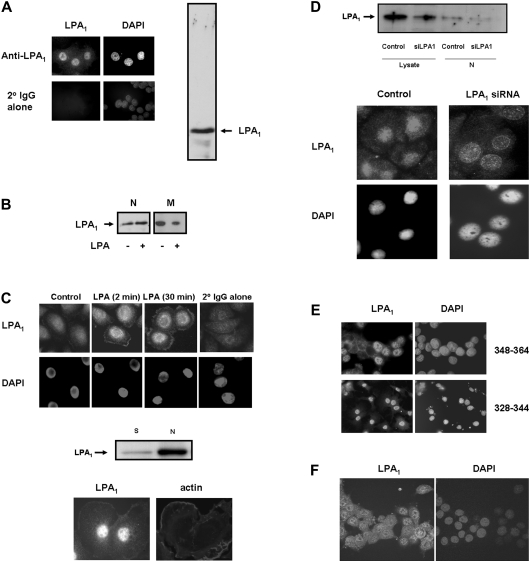
Figure 1. Detection of nuclear LPA1.
(A and B) Serum-deprived PC12 cells or (C) HBECs were immunostained with anti-LPA1328–344 antibody (A and B) and anti-(N-terminal LPA1) antibody (C), and detected using anti-(rabbit FITC-conjugated) secondary antibody. DAPI staining was used as a nuclear marker. (A) Western blot (right hand panel) was probed with anti-LPA1328–344 antibody, which showed a single immunoreactive band corresponding to LPA1 in the low-speed nuclear pellet from PC12 cells. (B) Western blot probed with anti-LPA1328–344 antibody showing the treatment of serum-deprived PC12 cells with 5 μM LPA for 10 min results in an increase in the amount of LPA1 in the low-speed nuclear pellet (N) and in a commensurate decrease in LPA1 in the high-speed membrane pellet fraction (M). (C) (Top panels) HBECs were treated with 1 μM LPA or untreated for the indicated times. (Bottom panels) PC12 cells were treated with 10 ng/ml NGF for 5 min to demonstrate plasma membrane localization of LPA1. PC12 cells and HBECs were immunostained with anti-LPA1328–344 antibody and anti-(N-terminal LPA1) antibody respectively. The middle panel shows a Western blot of high-speed hypotonic-treated nuclear pellets (N) and hypotonic/detergent high-speed supernatant fraction isolated from a nuclear pellet treated with this buffer mix (S) from HBECs, which was probed with anti-(N-terminal LPA1) antibody. (D) The upper panel shows a Western blot probed with anti-(N-terminal LPA1) antibody looking at the effect of siRNA-mediated elimination of the LPA1 in lysates and high-speed hypotonic-treated nuclear pellets (N) from serum-deprived HBECs. The lower panel shows immunofluorescence of control and siRNA LPA1-treated HBECs stained with anti-(N-terminal LPA1) antibody. (E) PC12 cells were immunostained with anti-LPA1 antibodies [and detected using anti-(rabbit FITC-conjugated) secondary antibody] raised against different C-terminal regions of LPA1 (see the Results section for details). DAPI staining was used as a nuclear marker. (F) Confocal immunofluorescent images of LPA1 nuclear localization in serum-deprived PC12 cells, using anti-LPA1348–364 antibody and detection with anti-(rabbit FITC-conjugated) secondary antibody.

