
Figure 2.
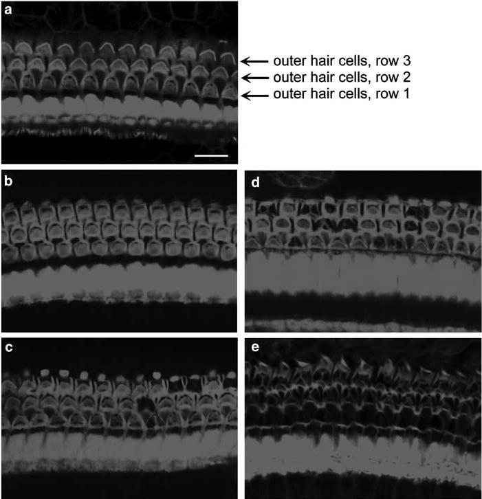
Hair cell loss in the cochlea. a: In the saline-treated CBA mice, rhodamine phalloidin staining for actin showed the well-defined outline of outer hair cells. b: After treatment with kanamycin for 7 days there was no apparent loss of outer hair cells. c: After treatment with kanamycin for 11 days some outer hair cells had disappeared in the basal turn. d: After 14 days of treatment with kanamycin, about 30% of outer hair cells were lost in the basal turn. e: At 1 week after the end of a 14-day treatment, almost all outer hair cells in the basal turn had vanished. The figure is representative of three individual animals at each time point. Scale bar: 10 μm