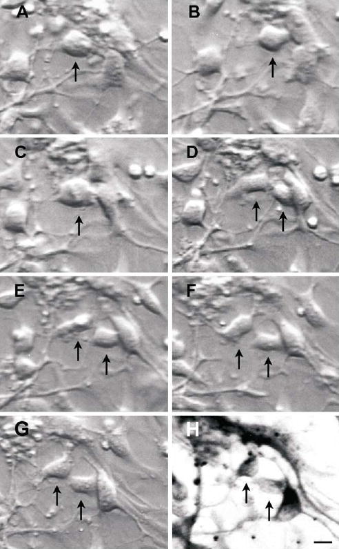
Figure 4.

Time-lapse photography of a dividing cell from a 9 week old foetus (↑ ), using differential contrast microscopy at 1h (B), 2.5 h (C) then at hourly intervals (D–G), over 6.5 hours, and the resultant daughter cells immunostained for β-III-tubulin (H). Note the relatively light β-III-tubulin staining, indicative of “new” neurons. Scale bar, 10 μm.
