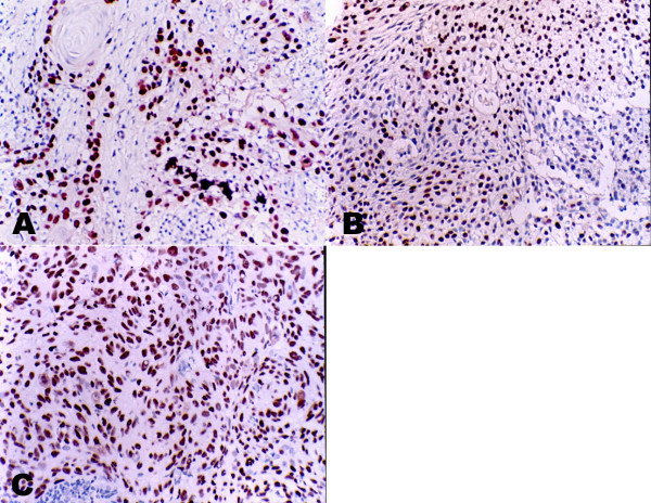
Figure 2.
Immunohistochemical demonstration of p63 protein expression in different histology NPC. Strong nuclear staining of p63 in keratinizing NPC (A) differentiated non-keratinizing NPC (B) and undifferentiated non-keratinizing NPC (C)(All the photomicrographs were taken in high-powered, ×400). The staining intensity is associated with the differential stage of NPC.

