
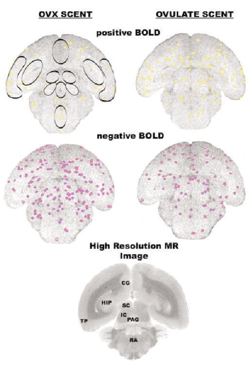
Figure 4.
Activational maps at the level of the midbrain. Images taken from a single animal. See Fig. 1 for detailed description. Abbreviations: SC = superior colliculus, IC = inferior colliculus, PAG = periaqueductal gray, RA = raphe.
Official websites use .gov
A
.gov website belongs to an official
government organization in the United States.
Secure .gov websites use HTTPS
A lock (
) or https:// means you've safely
connected to the .gov website. Share sensitive
information only on official, secure websites.
Activational maps at the level of the midbrain. Images taken from a single animal. See Fig. 1 for detailed description. Abbreviations: SC = superior colliculus, IC = inferior colliculus, PAG = periaqueductal gray, RA = raphe.