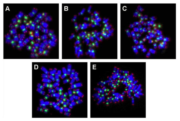
Figure 2.
PNA-FISH on metaphase spreads obtained from hESC exposed to (A) physiological control maintained at 37oC, (B) 4oC for 24h, (C) 25oC for 24h, (D) 4oC for 48h, (E) 25oC for 48h, The chromosomes were counterstained with DAPI (blue fluorescence). The telomere-specific PNA probe displayed red fluorescence, while the centromere-specific PNA probe displayed green fluorescence. In all experimental groups analyzed, there were no chromosomal aberrations.

