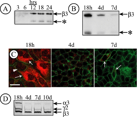
Figure 1.
LN5 is only expressed in MDCK cells at early times after plating. (A) Endogenous matrix proteins deposited by MDCK cells (8 × 10⁶ cells/10-cm2 culture dish) for the indicated times were immunoblotted with an antibody against the laminin β3 subunit. Immunoreactive material corresponding to deposited LN5 was detected beginning at 6 h plating, with increasing amounts afterward. An apparent β3 degradation product was also observed (*). (B) Immunodetection of endogenous matrix proteins deposited on the culture dish by MDCK cells 18 h, 4 d, or 7 d after plating. Under these conditions, LN5 deposition declines significantly after 4 d and is nearly absent after 7 d. As in A, a β3 degradation product is visible (*). (C) MDCK cells (7.5 × 10⁵ cells/35-mm dish) were plated on coverslips and fixed and stained with a polyclonal antibody against LN5 at either 18 h, 4 d, or 7 d after plating. Coverslips were observed by confocal fluorescence microscopy; only sections through the center of the cell are shown. After 18 h, LN5 staining (red) is apparent throughout the cytoplasm in a pattern resembling the endoplasmic reticulum. By 4 d, only a few cytoplasmic vesicles per cell are visible, with only isolated staining (arrows) by 7 d. Green, actin. Bar, 10 μm. (D) Pulse labeling and immunoprecipitation of LN5 in MDCK cells. MDCK cells (1 × 10⁶ cells/35-mm dish) were pulse labeled for 30 min at the indicated times after plating with 50 μCi of [35S]Met/Cys. After labeling, the cells were extracted with RIPA buffer, the extracts were immunoprecipitated with polyclonal anti-LN5, and the immunoprecipitates were analyzed by SDS-gel electrophoresis and fluorography. After 18 h plating, all three LN5 subunits are detected, whereas little α3 or γ2 is detected at later times.

