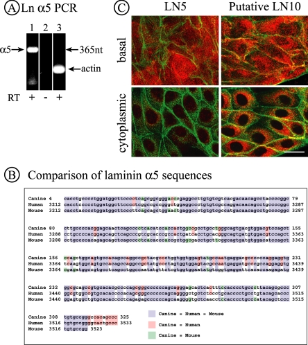
Figure 2.
MDCK cells synthesize the laminin α5 subunit. (A) PCR products corresponding to laminin α5 and β-actin were detected in RNA from MDCK cells by RT-PCR using primers specific to human laminin α5 and canine β-actin. Lanes 1 and 2, laminin α5 primers; lane 3, actin primers. (B) The PCR product shown in A was sequenced and the sequence compared with that of human and mouse laminin α5. The canine sequence is 85% identical to the human sequence and 81% identical to the mouse sequence. (C) MDCK cells (7.5 × 10⁶/35-mm dish) were plated for 42 h and then fixed and stained for either LN5 or putative LN10, the latter using a polyclonal antibody against LN1. Confocal sections from either the extreme base of the cells (top) or through the center of cells (bottom) are illustrated. In the case of LN5, significant amounts of deposited LN5 are visible at the base, but the cytoplasm has only limited amounts of perinuclear staining. For LN10, both the base and cytoplasmic compartments are strongly stained, indicating not only deposition of the protein but also continued synthesis. Bar, 20 μm.

