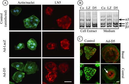
Figure 7.
Laminin β3 and γ2 are secreted after siRNA-mediated knockdown of laminin α3. (A) Control MDCK cells or cells infected for 18 h with Ad-LacZ or Ad-D5 were replated on uncoated glass coverslips for 24 h and stained with antibodies against LN5 (red), fluorescent phalloidin (green), and DAPI (blue), and viewed by conventional immunofluorescence microscopy. Anti-LN5 antibody staining is visible at the base in all three cases, although the staining pattern under cells expressing siRNA (Ad-D5) exhibits a characteristic “rose petal” pattern. Bar, 20 μm. (B) Control MDCK cells or cells infected with Ad-LacZ or Ad-D5 were metabolically labeled overnight. Detergent cell extracts and culture medium were immunoprecipitated with anti-LN5, and precipitated polypeptides were visualized on SDS-gels by fluorography. In extracts from Ad-D5–infected cells, the laminin α3 band is absent and laminin β3 and γ2 are increased in intensity (D5 lane). In the medium, laminin α3 is detectable but diminished, and the ratio of the β3 and γ2 bands to α3 is higher than controls, suggesting secretion of β3 and γ2 uncomplexed to α3 (compare D5 with Co and LZ). (C) Control MDCK cells or cells infected for 42 h with Ad-D5 were replated on uncoated glass coverslips for 24 h, stained with antibodies against LN5 (red) and fluorescent phalloidin (green), and viewed by confocal fluorescence microscopy. Individual optical sections taken at either the base of the cells or through the cell center are shown. Cells infected with Ad-D5 are exceedingly spread and still exhibit some staining with anti-LN5 at the base of the cell. Inside the same cells, anti-LN5 staining is intense in what seems to be the endoplasmic reticulum (arrow). Control (uninfected) cells seem normal with some anti-LN5 staining both within the cell and at the cell base. Ad-LacZ–infected cells seemed identical to uninfected controls (our unpublished data). Bar, 10 μm.

