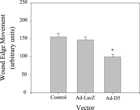
Figure 9.
Knockdown of laminin α3 slows wound-edge migration. Control MDCK cells or cells infected for 18 h with Ad-LacZ or Ad-D5 were replated at confluent density on gridded, uncoated glass coverslips for 24 h, and wounded by scraping. Wound-edge migration relative to the time of wounding was measured on micrographs after 24 h. Note that wound-edge movement occurred under all three conditions but was significantly reduced in cells expressing siRNA (Ad-D5). *p < 0.05.

