Figure 1.
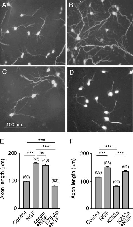
NGF induced axonal elongation is mediated via p75NTR in hippocampal neurons. Cells were plated onto glass coverslips at a density of 150 neurons/mm2. After 2 DIV, NGF or NGF plus the p75-antibody or NGF plus 1% rabbit neutral serum was added to the cells and incubated for a further 16 h. In parallel experiments, the Trk inhibitor K252a was added to the cells in the absence or presence of NGF and incubated for a further 16 h. Digital fluorescence images of phosphorylated neurofilament immunostaining by SMI31 mAb in control cultures (A), 100 ng/ml NGF (B), 100 ng/ml NGF plus p75-antibody antiserum, diluted 1:100 (C); or 200 nM K252a (D). The means, SE, and number of the neurons observed (in brackets) for each treatment are shown (E and F). Note that NGF induced axon elongation and the application of the anti-p75NTR blocking antibody prevented such an effect, whereas the application of the neutral rabbit serum did not (E). In contrast, K252a did not prevent the axon growth promoted by NGF, although, by itself, decreased axon length (F). Here and in the following figures, the levels of significance (asterisks) were determined for the data sets connected by horizontal lines using an unpaired t test. *p < 0.05, **p < 0.01, and ***p < 0.001.

