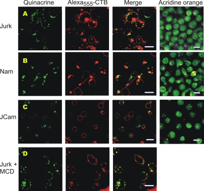
Figure 5.
Confocal microscopic analysis of putative ATP stores in lymphocytes. Live Jurkat T-cells (A), Namalwa B-cells (B), and lck-deficient Jurkat mutants J.CaM (C) were costained with cell surface marker Alexa555-CTB (red) and marker of putative ATP stores quinacrine (green), as well as with another acidotropic dye acridine orange. (D) CTB- and quinacrine-labeled Jurkat were pretreated for 30 min with 10 mM methyl-β-cyclodextrin (MCD) before microscopic inspection. Z-stack reconstruction of the adjacent images of Jurkat cells costained with Alexa555-CTB and quinacrine can also be viewed in movie format in the Supplementary Video. Bar, 10 μm.

