Figure 5.
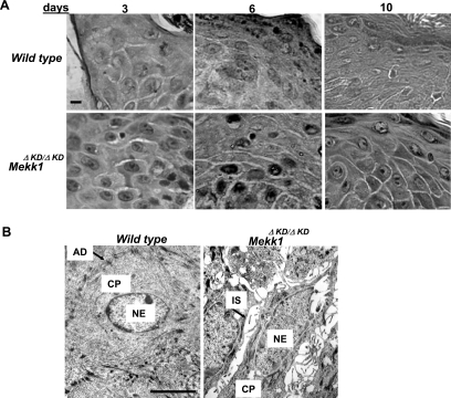
Lack of cell–cell junction formation in the wound epidermis of Mekk1ΔKD/ΔKD mice at late stage of healing. Morphological examination of the wounding edge epidermis at various days after injury. The wound tissues were examined under either phase-contrast microscopy (A) after H&E staining, and pictures were taken at 1000× magnification (bar, 10 μm), or electron microscopy (B), which was done on day 6 wounds. At early stage (day 3), both wild-type and Mekk1ΔKD/ΔKD mice show an apparent increase in intercellular spaces in the wounding edge epithelium. At late stage (days 6 and 10), only the Mekk1ΔKD/ΔKD mice display the spaces, whereas wild-type mice show tight cell–cell contact and the formation of desmosomes, similar to those present in normal unwounded skin. CP, cytoplasm; NE, nucleus. Bar, 10 μm.

