Figure 3.
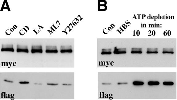
(A) Effects of various actin inhibitors on the total level of adhesive dimers. CD, cytochalasin D; LA, Latrunculin A; ML7, ML-7, Y27632, Y-27632; Con, control cells. Overnight AEcM/AEcF cocultures were treated by different inhibitors for 20 min and then were immunoprecipitated by an anti-myc antibody. The blots were probed for the presence of the immunoprecipitated Ec1M (myc) or coimmunoprecipitated Ec1F (flag). The latter could derive only from adhesive Ec1M-Ec1F dimers. (B) Effect of ATP depletion on the adhesive dimers. Overnight AEcM/AEcF cocultures were incubated in HBS buffer supplemented with glucose for 20 min (HBS), or in HBS buffer containing metabolic inhibitors for 10, 20, or 60 min (indicated above the lanes).

