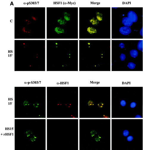
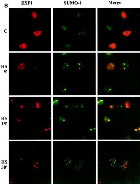
FIG. 9.
HSF1 and SUMO-1 transiently form cogranules at the onset of the heat shock response. (A) (Upper panel) HeLa cells were transiently transfected with HSF1-Myc and heat shocked for 15 min at 42°C or left untreated (C). Methanol-fixed cells were double stained by using anti-p-S303/7 antibody to detect phosphorylated HSF1 and anti-Myc antibody to localize total HSF1. Immunostaining was analyzed by fluorescence microscopy. Colocalization can be seen as yellow in the merged image. DAPI was used for nuclear staining. (Lower panel) HeLa cells were transiently transfected with HSF1-Myc and heat shocked for 15 min at 42°C. Cells were double stained with anti-p-S303/7 and anti-HSF1 antibodies in the presence or absence of 150 μg of recombinant human HSF1 (rHSF1) (17)/ml. (B) HeLa cells were transiently transfected with wild-type HSF1-Myc and GFP-SUMO-1 and heat shocked at 42°C for indicated times or left untreated (C). HSF1 was detected by using monoclonal anti-Myc antibody combined with a red fluorescent secondary antibody, and GFP-SUMO-1 was visualized through the green channel. (C) HeLa cells were transiently transfected with S303A or K298R HSF1 mutants together with GFP-SUMO-1. Cells were heat shocked at 42°C for 15 min and analyzed as described above.


