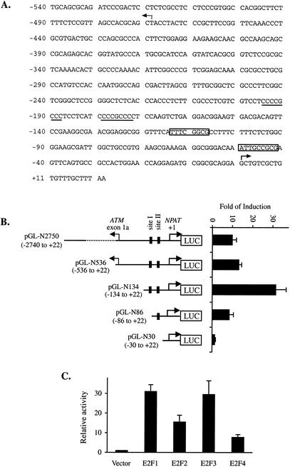
FIG. 2.
Activation of the NPAT promoter by E2F. (A) Sequence of the human NPAT promoter. The DNA sequence between the human ATM and NPAT genes is shown. The two putative E2F recognition sequences are boxed. Two potential SP1 sites are underlined. The +1 position for NPAT is indicated by the solid arrow. The broken arrow indicates the start site for the ATM gene (41). (B) Activation of the NPAT promoter by E2F1. A schematic representation of the NPAT promote-luciferase (LUC) reporter constructs is shown on the left. The nucleotide positions of the NPAT promoter fragments are indicated in parentheses. Site I and site II indicate the two E2F sites. U2OS cells were transfected with 1 μg of pCMV (vector) or 1 μg of pCMV-E2F1 together with 50 ng of the indicated NPAT promoter-luciferase reporter construct. For normalization of transfection efficiency among the different samples, 50 ng of pCMV-lacZ was also cotransfected. At 24 h after transfection, the cells were lysed and the activities of luciferase and β-galactosidase were assayed as described in Materials and Methods. Fold induction was calculated by comparing the normalized luciferase activity from E2F1-transfected cells with that from vector-transfected cells. The means and standard deviations from at least three independent experiments are shown. (C) Regulation of the NPAT promoter by E2F proteins. U2OS cells were transfected with 1 μg of pCMV (Vector) or 1 μg of the indicated E2F-expressing plasmid together with 50 ng of pGL-N134 and 50 ng of pCMV-lacZ. The samples were analyzed as described for panel B. The normalized luciferase activity from the cells transfected with the vector was set as 1.

