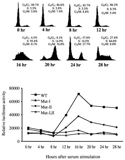
FIG. 6.
Activation of the NPAT promoter at the G1/S-phase boundary during growth stimulation depends on E2F recognition sites. NIH 3T3 cells were grown in six-well plates and transfected with 300 ng of wild-type (WT) or mutant (Mut) NPAT promoter constructs as indicated. For normalization of transfection efficiency, 200 ng of pCMV-lacZ was also cotransfected. At 18 h after transfection, the cells were washed and cultured in 0.1% serum. Cell proliferation was induced with 20% serum 48 h after serum starvation. At the indicated times, the cells were harvested and assayed for cell cycle distribution (top) and luciferase and β-galactosidase activities. The results shown are the means for triplicate samples. Similar results were also obtained in another independent experiment (data not shown).

