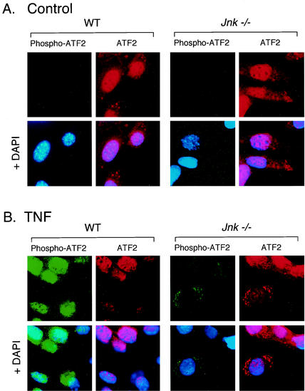
FIG. 5.
Effect of JNK deficiency on phosphorylation and subcellular location of ATF2. ATF2 and phospho-ATF2 were examined by immunofluorescence microscopy. Fibroblasts were treated without (A) and with (B) TNF-α (10 ng/ml for 15 min), fixed, and stained with antibodies to ATF2 (Texas red [red]) and phospho-ATF2 (FITC [green]). DNA was stained with DAPI (blue). The cells were imaged by conventional fluorescence microscopy.

