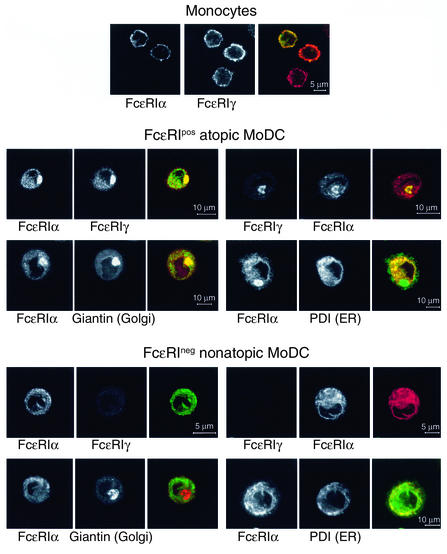
Figure 4.
Monocytes, FcεRIpos, and FcεRIneg DCs show a different localization of FcεRI subunits. Cells were adhered to coverslips, fixed, and permeabilized using 0.1% saponin. After blocking using human IgG Fc, sequential indirect immunolabeling with Ab against FcεRI subunits and organelle markers was performed. After mounting, the samples were analyzed by confocal laser scanning microscopy.

