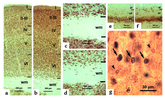
Figure 4.
Photomicrographs of NeuN-immunostained coronal sections of the primary somatosensory cortex (a–d) and hippocampal CA1 (e and f) in LID-plus-KI (a, c, and e) and LID-1 (b, d, and f) progeny at P40. In the neocortex of LID-1 pups, borders between layers are more blurred (horizontal dashed lines in b) than in LID-plus-KI rats (horizontal lines in a). The number of NeuN-labeled neurons increases both in subcortical white matter and in strata oriens and alveus of hippocampal CA1 of LID-1 rats (d and f, respectively) as compared with LID-plus-KI rats (c and e). In g, CNP-positive oligodendrocytes (arrowheads) and BrdU-labeled nuclei (arrows) are shown in layer V of a LID-1 rat from an E14–E16 subgroup. Note that CNP-positive oligodendrocytes are BrdU negative.

