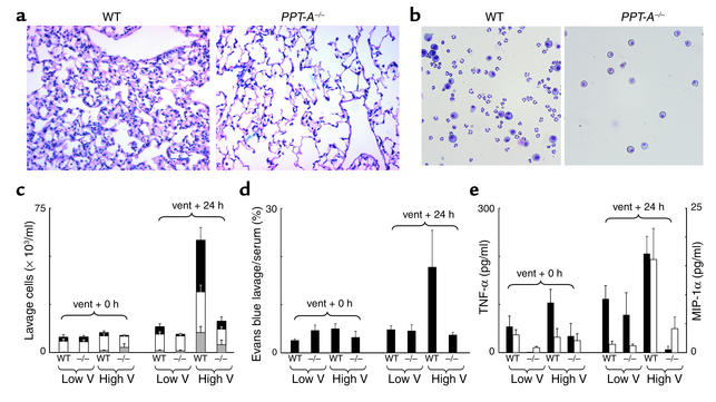
Figure 2.
Protection conferred against stretch-mediated injury by PPT-A gene deletion. (a) H&E-stained sections (×20) obtained 24 hours after a 4-hour period of mechanical ventilation with a tidal volume of 20 ml/kg demonstrate edema and inflammatory infiltration of the pulmonary interstitium in WT mouse but not in PPT-A gene–deleted (PPT-A–/–) mouse. (b) Microphotograph (×40) of bronchoalveolar lavage fluid after similar treatment shows exudation of macrophages and neutrophils in WT mouse, but only small numbers of macrophages in PPT-A–/– mouse. (c) Cell counts in the bronchoalveolar lavage fluid (n = 10 for each combination of variables)show increased numbers of neutrophils (gray bars), macrophages (white bars), and erythrocytes (black bars) in WT but not in PPT-A–/– mice 24 hours after ventilation (vent + 24 h) with high tidal volumes (High V) (P < 0.0001). (d) Evans blue lavage/serum ratios (n = 10)demonstrate increased alveolar-capillary permeability in WT but not in PPT-A–/– mice 24 hours after High V (P < 0.002). (e) TNF-α levels (black bars; n = 5) were higher in WT than in PPT-A–/– mice after ventilation with High V, both at the end of the 4-hour ventilation period (vent + 0 h; P = 0.02) and 24 hours later (P < 0.0001). MIP-1α levels (white bars; n = 5) were higher in WT than in PPT-A–/– mice after High V, but only after 24 hours (P < 0.0001). Values are shown as mean ± SE.

