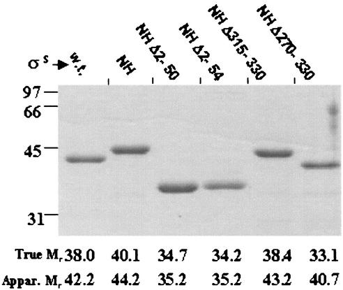
FIG. 2.
Electrophoretic migration of σS and its derivatives. The indicated σS derivatives were electrophoresed (37) on a 10% polyacrylamide gel with sodium dodecyl sulfate and visualized after staining with Coomassie blue. At left are shown the positions of migration of marker proteins of indicated Mr. For each σS derivative, the true and apparent (Appar.) Mr values, the latter as calculated from the migration rate on the gel, are given below the corresponding lane. w.t., wild-type (native) σS.

