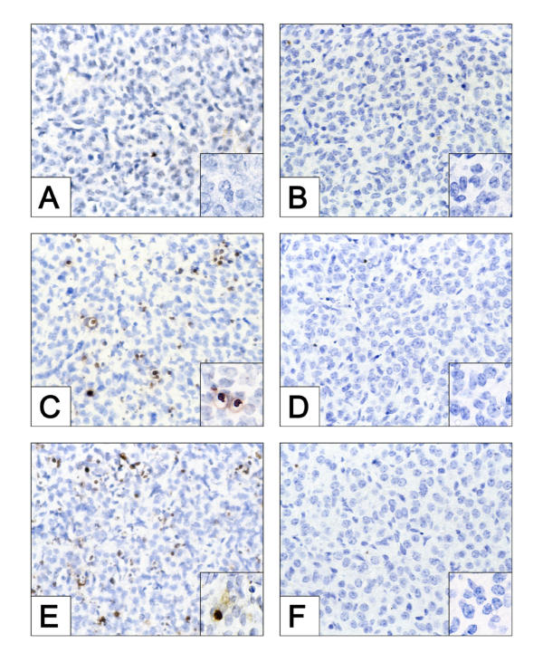
Figure 1.
Histochemical assessment of apoptosis in CL derived from WT and caspase-3-null mice following treatment with IgG, Jo2 or PGF2α, each administered at 48 h post-ovulation. The ovaries were harvested 8 h post-injection. The data shown depict the incidence of apoptotic (TUNEL-positive, brown staining) cells in CL of ovaries derived from WT (A, C, and E) and caspase-3 deficient mice (B, D, and F) following injection with IgG (A, B), Jo2 (C, D) or PGF2α (E, F). Original magnifications, × 600. The insets represent a higher magnification (× 1000), demonstrating the presence or absence of TUNEL positive cells. The photomicrographs shown are representative of similar results obtained in at least three independent experiments.

