Figure 3.
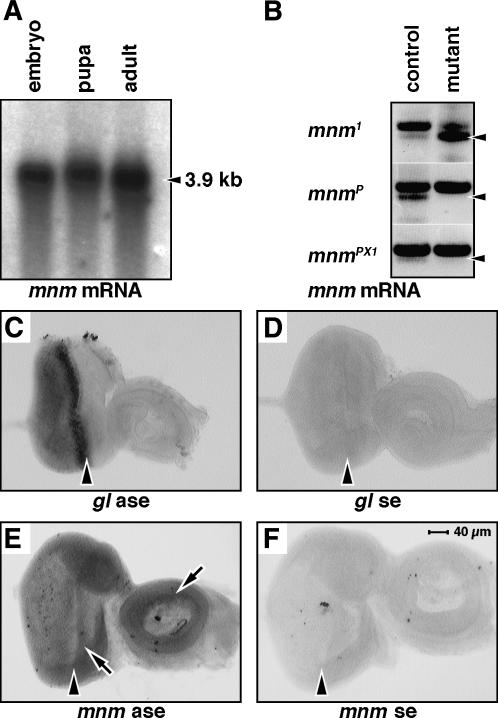
mnm expression. (A) RNA gel blot, stages as indicated. mnm mRNA is indicated by an arrowhead. (B) RT gel. RNA from single embryos was isolated and analyzed by RT–PCR using gene-specific mnm primers. The arrowheads mark the predicted RT product in all sections. The top band in all sections is the predicted product from genomic DNA remaining in the reaction. Genotypes are mnm1/CyO and mnm1 (top), mnmP/CyO and mnmP (middle), and mnmPX1/CyO and mnmPX1 (bottom). There was no detectable transcript in mnmP or mnmPX1 homozygote embryos compared to the heterozygote controls (middle and bottom sections). The mnm transcript appeared to be overexpressed in the mnm1 homozygote embryos compared to controls (top), suggesting that mnm1 may be a hypermorphic or neomorphic allele. (C–F) RNA in situ hybridization experiments: (C) glass antisense, (D) glass sense, (E) mnm antisense, and (F) mnm sense. Third instar eye-imaginal discs: anterior right, to the same scale indicated in F. The morphogenetic furrow is indicated by an arrowhead. Note the elevated expression of mnm mRNA in the eye field and in parts of the antenna (arrows in E).

