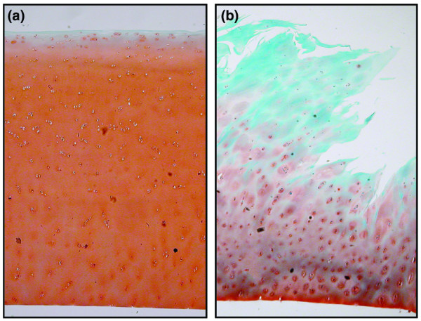
Figure 1.

Normal healthy and osteoarthritic cartilage histology. Representive light micrographs of condylar cartilage obtained post mortem from joints with (a) normal healthy cartilage and (b) cartilage obtained at joint replacement surgery. Sections are stained with safranin-O fast green-iron haematoxylin and graded for features of osteoarthritis according to the slightly modified criteria [23] described by Mankin and coworkers [24]; scores for the shown samples are 0 and 7, respectively.
