
Figure 5.
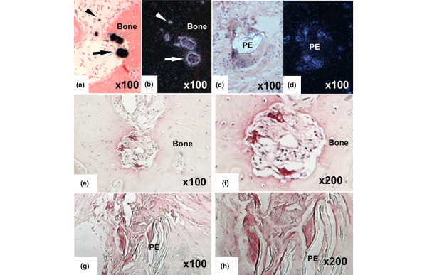
Detection of TRAP mRNA and TRAP enzymatic activity in sections of human peri-implant tissues. The techniques used were in situ hybridization with a 35S-labeled antisense RNA probe ((a, c) bright field and (b, d) and dark field) and (e-h) histochemistry. TRAP mRNA expression is detected in mononuclear (arrows) and multinuclear cells (arrowheads) on the bone surface adjacent to the peri-implant granuloma (panels a and b). Cells associated with polyethylene (PE) particles also express TRAP mRNA (panels c and d). The enzymatic activity is evident as purple staining seen in similar cells (panels e-h). TRAP, tartrate-resistant acid phosphatase.