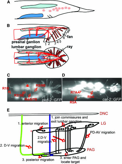
Figure 1.—
Ray axon pathways in wild type. (A) Lateral view of the L4 larval male tail, showing the positions of the nine neurons of one type before they migrate into the lumbar ganglion during remodeling of the tail. (B) Lateral (top) and ventral (bottom) views of the adult male tail, showing the fan and rays and one of the two sets of ray sensory neurons. Ray neuron sensory dendrites extend to the tips of the rays, cell bodies are in the lumbar ganglia, and axonal output is in the pre-anal ganglion. (C and D) Fluorescence photomicrographs of strains carrying pkd-2∷GFP and cat-2∷GFP. pkd-2∷GFP labels all RnB neurons except R6B; cat-2∷GFP labels R5A, R7A, and R9A. In C, commissural pathways are indicated; the open arrow indicates a region of diffuse fluorescence within the pre-anal ganglion where there is a concentration of ray synapses. (E) The ray axons follow various pathways between the lumbar ganglia and the pre-anal ganglion. The axon of R1B follows a segmented pathway defined by apparent choice points (labeled *), first migrating anteriorly out of the pre-anal ganglion, then turning to traverse around the body wall to the ventral side, then progressing ventrally into the pre-anal ganglion. On the right side, there are two ventrodorsal commissures in the vicinity of the ray 1 commissure, one containing AS11 (blue) and a second containing motor neurons VD12 and VD13 (green). R1BR axon could follow one of these or pioneer its own route. The axon of R2B sometimes follows a similar pathway, but more often fasciculates with the axon of R3B in a dorsoventral commissure. The axons of R4B, 5B, 7B, and presumably 6B follow a similar dorsoventral commissure. The axons of R8B and R9B follow the preexisting lumbar commissure. RnA neurons follow similar pathways and can fasciculate with the RnB neurons. For a diagram of these pathways in transverse section showing the pathway of the ray axon commissures between the basal surface of the hypodermis and the hypodermal basal lamina, see Figure 7.

