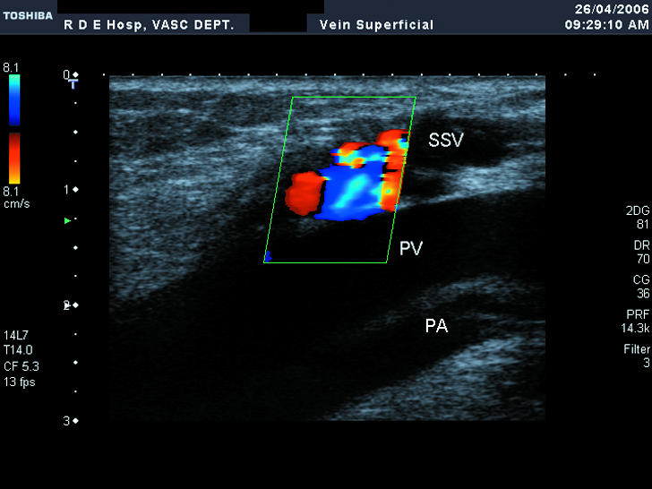
Fig 3.

Duplex ultrasound scan of varicose veins showing the short saphenous vein (SSV) joining the popliteal vein (PV) with the popliteal artery (PA) adjacent. The patient is standing, and the calf has just been squeezed and released: the colour indicates reflux down the short saphenous vein as a result of an incompetent valve at the saphenopopliteal junction
