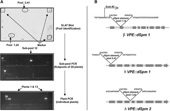
Figure 2.
Identification, Isolation, and Location of dSpm Transposon Insertions in δVPE and βVPE.
(A) The top gel shows a chemiluminescence image resulting from hybridizing a SLAT blot with a δVPE probe to identify parental pools containing dSpm insertions (pools 5.41 and 1.24) in or near the δVPE gene. The middle gel shows PCR identification (see Methods) of a subpool of progeny containing an insertion within δVPE (pool 5.41), and the bottom gel shows PCR identification of two individual plants carrying this insertion.
(B) Illustrations showing the locations of dSpm insertions within the δVPE and βVPE genes with respect to DNA sequence and intron/exon regions. Gray boxes indicate exon (coding) regions of the genes.

