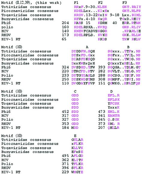
Table 1. Conserved motifs in the viral RdRps and HIV-1 reverse transcriptase (for comparison).
The first amino acid of each motif and/or region is indicated to the left of the sequence. Sequences and numbers are according to the PDB files when GenBank and PDB files differ. Distances between motifs are similar in all the RdRps, as shown in Figure 1 (with the exception of distances within motif F, see text). Similar or identical residues are in pink.

