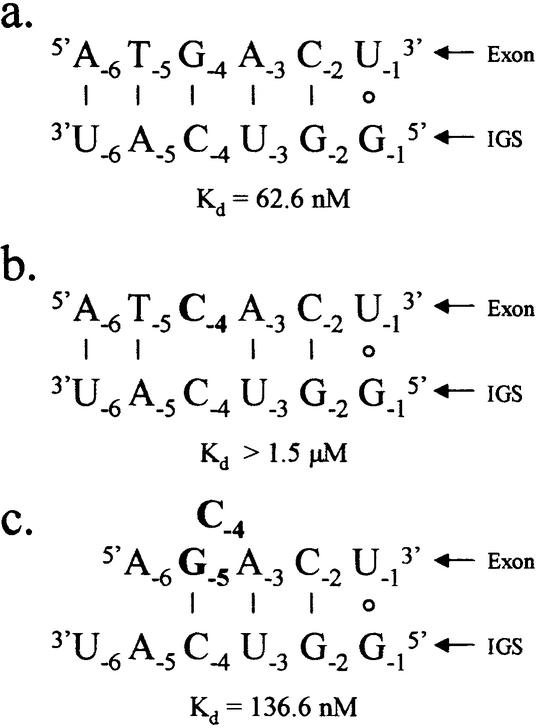
Figure 5.
Schematics of 5′ exon mimics base pairing with the IGS of the P.carinii ribozyme. (a) The native 5′ exon, (b) the 5′ exon with a single G to C mutation at exon position –4 and (c) the 5′ exon with a G to C mutation at exon position –4 and a T to G mutation at exon position –5. The Kd values given are for the respective 5′ exon mimics binding to the IGS through base pairing and tertiary interactions (in 15 mM MgCl2). Exon positions –2 through to –6 are deoxyribonucleotides. Note that the double mutant (c) rescues binding lost with the single mutant (b).

