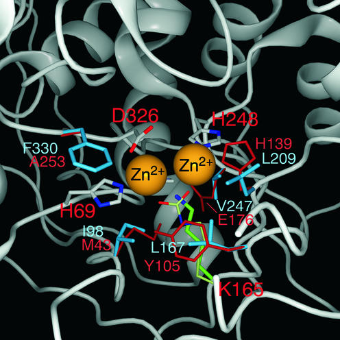
Figure 4.
Modeling of the human DHPase structure onto the known E.coli DHOase structure. The figure shows a close-up of the active site with amino acids within a 7.5 Å distance from the zinc ions. Amino acid side chains coordinating to the zinc ions (H69, K165, H248 and D326) are shown in the normal atomic colors. H67 is behind one of the zinc atoms and is therefore not visible in this figure. With the exception of the non-carboxylated K165 all other residues are in perfect overlap with the template structure of DHOase. The side chains of all other amino acids of the DHPase active center are in cyan, whereas the residues of template DHOase are in orange-red.

