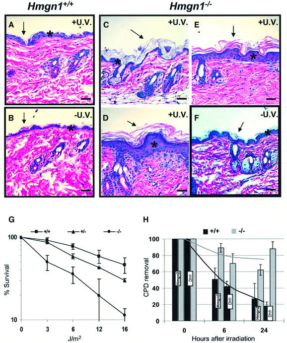
Fig. 2. Loss of HMGN1 leads to UV hypersensitivity. (A–F) Increased sensitivity of Hmgn1–/– mice to UV-B. Histology of skin after cumulative irradiation of 1200 J/m2 UV-B. (A and B) Hmgn1+/+ mice, (C–F) Hmgn1–/– mice. Note the increased acanthosis (asterisk; compare E and D with F) and hyperkeratosis (arrows) in the epidermis of the Hmgn1–/– mice. (G and H) Impaired UV repair in MEFs lacking HMGN1. (G) Increased UV sensitivity of Hmgn1–/– fibroblasts. Shown is survival 72 h after irradiation with the indicated doses (see Materials and methods). (H) Decreased rate of gene-specific CPD removal in Hmgn1–/– fibroblasts. Shown is the quantitative analysis of Southern blots with probes specific for the Dhfr and Hmgn2 genes as described in Materials and methods. The 0 h point represents the initial lesion frequency. The bar graphs represent the average of three experiments.

An official website of the United States government
Here's how you know
Official websites use .gov
A
.gov website belongs to an official
government organization in the United States.
Secure .gov websites use HTTPS
A lock (
) or https:// means you've safely
connected to the .gov website. Share sensitive
information only on official, secure websites.
