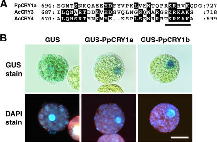
Figure 2.
Intracellular Distribution of GUS-PpCRY Fusion Proteins in Moss Protoplasts.
(A) Alignment of amino acid sequences of the C termini of PpCRY1a, AcCRY3, and AcCRY4. The conserved amino acid residues are highlighted. The putative nuclear localization signal is underlined.
(B) Protoplasts incubated under white light. The top row shows GUS activity, and the bottom row shows the positions of the nuclei in the same cells via 4′,6-diamidino-2-phenylindole (DAPI) fluorescence. Bar = 20 μm for all panels.

