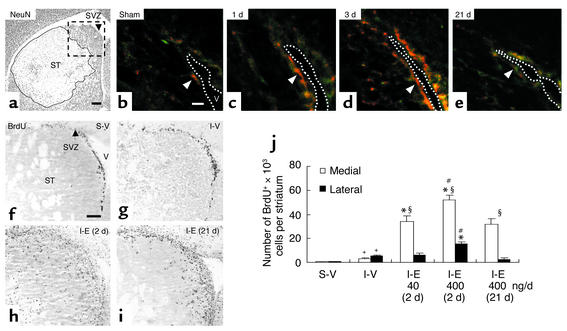
Figure 1.
EGF-enhanced proliferation after ischemia. (a) Ischemia reperfusion caused a marked loss of NeuN+ neurons (outlined area) confined to the striatum (ST). SVZ, subventricular zone. Scale bar is 200 μm. (b–e) Without ischemia (b), few nestin+ cells (green) expressing EGF receptors (red) surrounded the lateral ventricle (V). After ischemia, the intensity of EGF receptor immunoreactivity in nestin+ SVZ cells (arrowheads) and the number of nestin+ cells expressing EGF receptor increased at 1 day (c), reached maximum at 3 days (d), and returned to basal levels at 21 days (e). The dotted line delineates lateral ventricle. Scale bar is 20 μm (n = 3–4/time-point). (f–i) BrdU+ cells at 9 days after sham or ischemia in an area corresponding to the box (a). After vehicle, the number of BrdU+ cells was higher after ischemia (g) than sham (f) (quantification in j). Ischemia followed by EGF (I-E; 400 ng/day) increased BrdU+ cells and was greater when EGF infusion started 2 days (h) rather than 21 days (i) after ischemia. (f) Scale bar is 100 μm. Bregma, 0.8 mm. (j) Quantification. S-V, sham-operated; I-V, ischemic group with vehicle; I-E (2d), ischemic groups with EGF (400 ng/day) initiated at day 2; I-E (21d), day 21 after ischemia. The effects of EGF (40 and 400 ng/day) initiated at day 2 were compared. +Significant difference between S-V and I-V; *between vehicle- and EGF-treated ischemic groups; #between groups treated with EGF 2 days or 21 days; §between medial and lateral striatum within the same group; n = 3–4/group, unpaired Student t test, P < 0.05.

