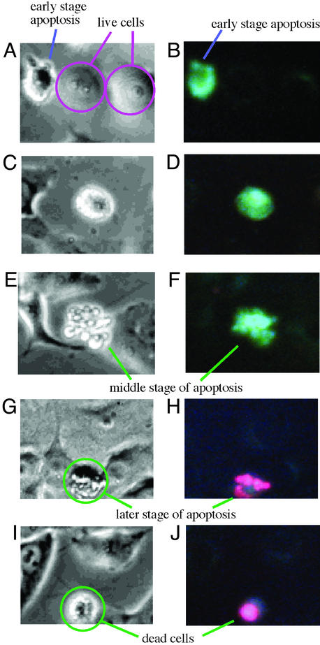
Figure 3.
Morphological changes of HeLa cells (dually labeled with 4 and PI) by simultaneous phase contrast (A, C, E, G, and I) and fluorescence (B, D, F, H, and J) microscopy (×400). (A and B) Live cells and the early stages of apoptotic cells. (C and D) Early stage of apoptosis. (E and F) Middle stages of apoptosis. (G and H) Later stages of apoptosis. (I and J) Dead cells.

