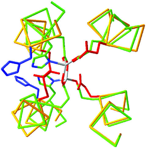
Fig 5.
Superposition of the di-Zn(II) and apo structures of DF1. The backbone trace of the di-Zn(II) structure is in green, and the side chains are plotted with Corey–Pauling–Koltun colors (C, green; N, blue; O, red). The backbone of apo-DF1 is in yellow, the Glu side chain is in red, and the His residue is in blue. (Right) Helices 1 and 1′, which superpose well between the structures. (Left) A much poorer superposition of helices 2 and 2′ arises from a rotation of the helices about their axes, which increases the exposure of the His and Glu side chains.

