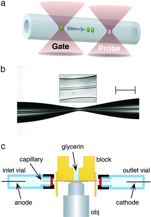
Figure 1.
(a) Schematic of the photoreaction and probe regions in optically gated microsecond separations. Femtoliter reaction plugs are created in flowing reagent streams by a gate focus that is switched to high intensity for ≈1–2 μs; transient reaction products migrate according to charge-to-drag ratios, providing the capacity to analyze reaction mixtures within microseconds. As depicted, photoproduct diffusion is minor on the time scales of these separations. (b) A typical hourglass structure created in the central portion of a short fused-silica capillary (29-μm i.d. and 320-μm o.d. in unpulled regions). By reducing the cross-sectional area by a factor of ≈30–40, fields in excess of 0.1 MV⋅cm−1 can be generated in short capillary stretches by using an applied potential of 20 kV. (Scale bar, 250 μm.) The boxed inset shows the central (≈60-μm) region of the hourglass where separations are performed. (c) Schematic of the electrophoresis assembly. Index-matching glycerin is used to fill microscopic gaps between the capillary and the underlying coverslip. The microscope objective (obj) focuses two separate laser beams, a microsecond-gated photoreaction beam and a continuous probe beam, to positions in the capillary separated by ≈10 μm.

