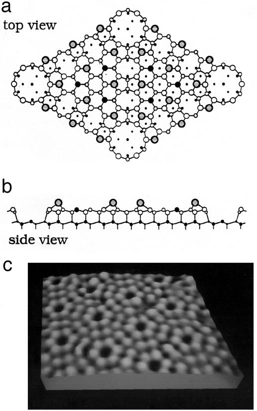
Figure 6.
The (7 × 7) structure of the (111) surface of silicon. (a) Schematic top view. (b) Schematic side plan view. (c) STM micrograph. The side view is a plan view of the nearest neighbor bonding in a plane normal to the surface containing the long diagonal of the surface unit cell. In the top view (a) the large shaded circles designate the adatoms in the top layer of the structure. It is evident from c that the STM images only these atomic species. The large solid circles designate second-layer “rest atoms” that are not bonded to an adatom. Large open circles designate triply bonded atoms in this layer. Small open circles designated 4-fold-bonded atoms in the bilayer beneath. Smaller solid circles designate atoms in the fourth and fifth bilayers beneath the surface. The size of circles is proportional to the distance from the surface. In the side view (b) smaller circles indicate atoms out of the plane of this diagonal. [(a and b) adapted from Takayanagi et al. (61) with permission; (c) after Joel Kubby, personal communication.]

