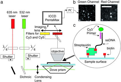
Figure 1.
The experimental system. (a) Schematic drawing of the optical setup. The green laser illuminates the surface in total internal reflection mode while the red laser is blocked. Both Cy3 and Cy5 fluorescence spectra are recorded independently by the intensified charge-coupled device. (b) Single-molecule images obtained by the system. The two images show colocation of Cy3- and Cy5-labeled nucleotides in the same template. (Scale bar = 10 μm.) (c) Schematic of primed DNA template attached to the surface of a microscope slide via streptavidin-biotin.

