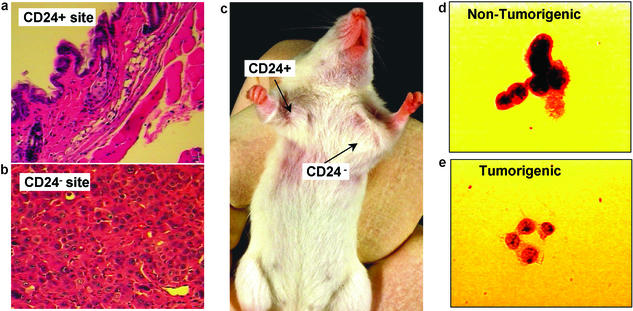
Figure 3.
Histology from the CD24+ injection site (a; ×20 objective magnification) revealed only normal mouse tissue, whereas the CD24−/low injection site (b; ×40 objective magnification) contained malignant cells. (c) A representative tumor in a mouse at the CD44+CD24−/lowLineage− injection site, but not at the CD44+CD24+Lineage− injection site. T3 cells were stained with Papanicolaou stain and examined microscopically (×100 objective). Both the nontumorigenic (d) and tumorigenic (e) populations contained cells with a neoplastic appearance, with large nuclei and prominent nucleoli.

