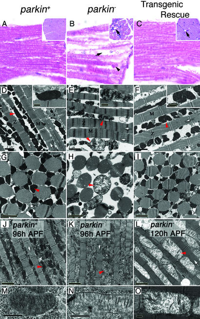
Figure 4.
Parkin mutants manifest muscle degeneration and mitochondrial pathology. (A–C) Hematoxylin and eosin staining of longitudinal sections of IFMs show well preserved muscle in controls (A), contrasted with acute degeneration in parkin mutants (B) characterized by vacuole formation (arrow) and accumulation of cellular debris (arrowhead). Transgenic expression of parkin substantially restores muscle integrity (C), though occasional vacuoles are still seen (arrow). (Insets) Transverse section of IFMs. (D and G) Sections through parkin+ adult IFMs show a regular and compact myofibril arrangement (white arrows) with many electron-dense mitochondria (red arrowheads and Inset). (E and H) parkin− adult IFMs show an irregular and dispersed myofibrillar arrangement with diffuse Z-lines and M-bands. Mitochondria are grossly swollen and malformed showing disintegration of cristae (red arrowheads and Inset). (F and I) Myofibril and mitochondrial integrity can be restored by transgenic expression of parkin in muscle tissue. (J–O) The mitochondrial pathology is progressive and precedes myofibril degeneration. (J and M) IFMs from control 96-h pupae show many electron-dense mitochondria, whereas age-matched parkin mutants (K and N) already have mitochondria that are less electron-dense showing fewer cristae. (L and O) By 120 h, parkin− pupae still show intact myofibril structure, but the mitochondria are profusely swollen as the cristae continue to degenerate. Genotypes: parkin+: w; parkrvA/Df(3L)Pc-MK, parkin−: w; park25/Df(3L)Pc-MK, transgenic rescue: w; UAS-park; park25/24B-GAL4, park25. (Scale bars: D–L, 2 μm; M–O and Insets in D–F, 0.5 μm.) Z, Z-lines; M, M-bands; APF, after puparium formation.

