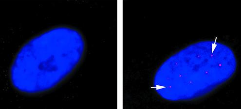
Figure 7.
Restoration of gems in SMA patient fibroblasts. Images show untransfected (a) and transfected (b) SMA type I fibroblasts. 4′,6-Diamidino-2-phenylindole staining highlights the nuclei in blue, and the white arrows indicate the gems (red dots in nucleus). Untransfected cells show 2–3% of their nuclei containing gems, whereas transfected cells show 13% gem-positive nuclei.

