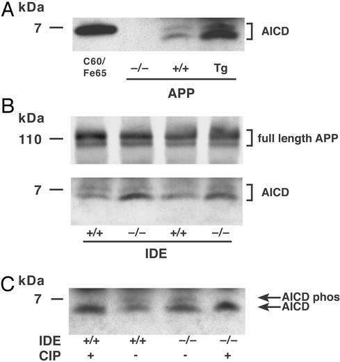
Figure 2.
Cerebral levels of APP and AICD in IDE-deficient mice. Shown are immunoprecipitation immunoblots of brain lysate with antibody C8. Identical results were obtained with C8 immunoprecipitation followed by anti-0443 immunoblotting. (A) AICD in APP gene-disrupted (−/−), WT (+/+), and APP transgenic (Tg) mouse brains. Lane C60/Fe65, the positive control, is lysate from transfected COS cells coexpressing a 59-aa AICD construct, which runs slightly higher than the endogenous 50-aa AICD, and Fe65, which stabilizes AICD (28). (B) Full-length APP and AICD in IDE −/− vs. +/+ brain. The APP and AICD bands are from identical lanes of the same blot, but the APP bands are from a lighter exposure. (C) Dephosphorylation of AICD. After the final wash following immunoprecipitation, samples were incubated for 1 h with either calf intestinal phosphatase (CIP) or buffer alone.

