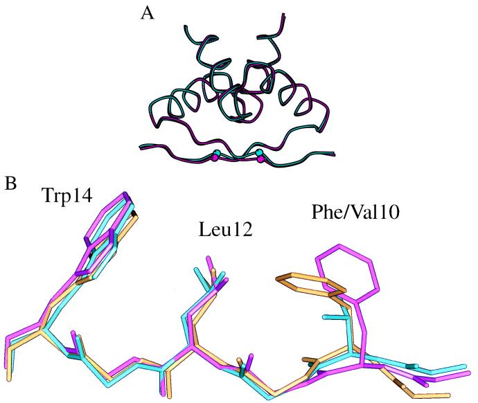
Figure 3.
(A) Superposition of backbone traces of the structures of the wild-type Arc dimer (cyan) and the mutant FV10 dimer (magenta). The Cα atoms of Phe-10 and Val-10 are shown as small spheres. (B) Superposition of the polypeptide backbones of residues 8–14 of wild-type Arc (magenta), the FV10 mutant (cyan), and wild-type Arc in the operator-bound complex (gold). The side chains of Phe-10 or Val-10, Leu-12, and Trp-14 also are shown.

