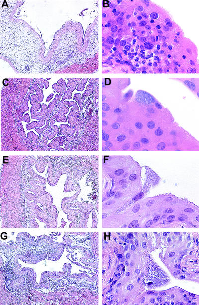
Figure 2.
Histology of infected bone marrow chimera bladders. Low- and high-power magnifications of infected bladder tissue from chimeric mice are shown. (Magnification: A, C, E, and G, ×5; B, D, F, and H, ×60.) Lpsn→Lpsn mice after infection have edema (A) and lymphocytic infiltrates (A and B). Infected Lpsd→Lpsd mouse bladders show minimal pathology (C) and infiltrating lymphocytes (C and D). Infected Lpsd→Lpsn mouse bladders have similar histology to Lpsd→Lpsd mouse bladders (E and F). Bladders from infected Lpsn→Lpsd mice show some edema (G) and lymphocytic infiltration (H).

