Figure 3.
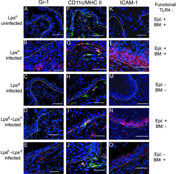
Fluorescent immunohistochemical analysis of infected bone marrow chimera bladders. Immunofluorescent analysis of bladder tissue is shown. (A–E) Gr-1 (Ly-6G) expression by neutrophils is shown in red. (F–J) CD11c (DC marker) staining is shown in red, and MHC II (macrophage/activated DC marker) is shown in green; coexpression of CD11c and MHC II by activated DCs appears in orange. (K–O) ICAM-1 expression by activated epithelium is shown in red. Bladder tissue was analyzed from uninfected Lpsn (A, F, and K), UPEC-infected Lpsn (B, G, and L), infected Lpsd (C, H, and M), Lpsd→Lpsn (D, I, and N), and Lpsn→Lpsd mice (E, J, and O). Nuclear morphology in all fields is shown in blue. The dashed line represents the basement membrane. Expression of functional TLR4 on the epithelial/stromal compartments (Epi) or on the bone marrow-derived compartment (BM) is indicated on the right. Neutrophils are present in UPEC-infected Lpsn (B) and, to a lesser degree, in infected Lpsn→Lpsd mice (E). (D) Occasionally, neutrophils could be seen in infected Lpsd→Lpsn mice. (F–J) DCs (red), macrophages (green), and activated DCs (orange) could be seen in all mice regardless of infection status. ICAM-1 expression could be clearly seen when infected mice express functional TLR4 on the epithelium [Lpsn (L) and Lpsd→Lpsn (N) mice]. (O) Weak ICAM-1 expression could be detected in the infected Lpsn→Lpsd mice.

