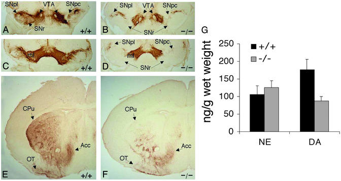
Figure 5.
TH immunohistochemistry of newborn (P2) wt (+/+) and ak/ak (−/−) ventral midbrain (A–D) and striata (E and F). Coronal sections through rostral (A and B) and middle (C and D) levels of midbrain were stained. TH IR was strong in the VTA of both wt and mutant mice but absent in mutant SNpc and SNr. ml, medial lemniscus. (E and F) Coronal sections throughout the striata of wt and ak mice were stained, and representative sections are presented. TH IR was reduced significantly in the dorsolateral region of mutant CPu when compared with wt striatum. (G) NE and DA levels in dissected P2 forebrains of wt and ak mice were measured by using HPLC. The data are presented as nanograms of neurotransmitter per gram of dissected tissue. The reduction of DA levels in mutant forebrains is statistically significant (n = 8, P < 0.001).

