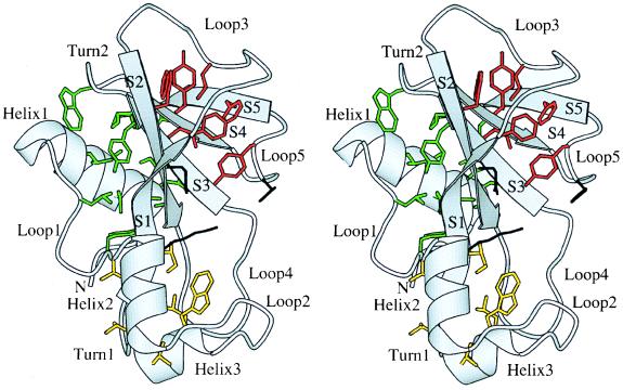
Figure 1.
Overall view of the structure of barnase (3). Secondary structure elements have been labeled. The residues belonging to the hydrophobic cores are shown in green (core 1), yellow (core 2), and red (core 3). The active site residues Lys-27, Glu-73, and His-102 are shown in black. This figure and all subsequent figures have been drawn with the program Bobscript (25).

