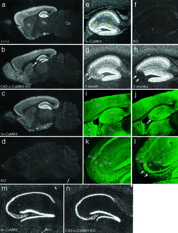
Figure 2.
Expression studies of CA3 α-CaMKII KO mice. (a–d) Dark field images of sagittal brain sections from 4-mo-old wild-type (a), α-CaMKII floxed (c), CA3 α-CaMKII KO (b), and global α-CaMKII KO (d) mice hybridized with an α-CaMKII-specific probe. Deletion of the fα-CaMKII gene was specific to hippocampal CA3. Cx, cortex; St, striatum; Th, thalamus; Cb, cerebellum. Arrow in b indicates the location of hippocampal CA3. (e–h) Dark-field higher magnification images of the hippocampus from α-CaMKII floxed (e), global α-CaMKII KO (f), and 1- (g) and 3-mo-old (h) CA3 α-CaMKII KO mice after hybridization with a probe specific for α-CaMKII. These images show that gene knockout is highly specific to the CA3 region of the hippocampus, indicated with double arrows. DG, dentate gyrus. (i–l) Confocal images from 3-mo-old floxed (i and k) and 4-mo-old CA3 α-CaMKII KO (j and l) mice after immunohistochemical staining with anti-α-CaMKII antibody. Lower-magnification sagittal images on top, followed by high-magnification images of hippocampal CA3 show that the α-CaMKII protein is absent from the vast majority of pyramidal cell bodies (stratum pyramidale; indicated with arrows) and dendrites (strata radiatum, oriens) in the CA3 region, whereas axons projecting from dentate granule cells to the stratum lucidum are strongly stained. H, hippocampus; sp, stratum pyramidale; sr, stratum radiatum; so, stratum oriens; sl, stratum lucidum; sl-m., stratum lacunosum-moleculare; GL, granule layer; ML, molecular layer. (m and n) Dark-field images of the hippocampus from 4-mo-old floxed (m) and CA3 α-CaMKII KO mice (n) after hybridization with a probe specific for β-CaMKII. No compensatory increase in β-CaMKII expression was observed in hippocampal regions.

